Priority Nursing Diagnosis for Hepatitis

Nursing Care Plan for Hepatitis

Nursing Care Plan for Hepatitis

Hepatitis is swelling and inflammation of the liver. It is not a condition, but is often used to refer to a viral infection of the liver. Characterized by the presence of inflammatory cells in the tissue of the organ.

Hepatitis may start and get better quickly (acute hepatitis), or cause long-term disease (chronic hepatitis). In some instances, it may lead to liver damage, liver failure, or even liver cancer.

How severe hepatitis is depends on many factors, including the cause of the liver damage and any illnesses you have. Hepatitis A, for example, is usually short-term and does not lead to chronic liver problems.

The symptoms of hepatitis include:
  •     Abdominal pain or distention
  •     Breast development in males
  •     Dark urine and pale or clay-colored stools
  •     Fatigue
  •     Fever, usually low-grade
  •     General itching
  •     Jaundice (yellowing of the skin or eyes)
  •     Loss of appetite
  •     Nausea and vomiting
  •     Weight loss
Many people with hepatitis B or C do not have symptoms when they are first infected. They can still develop liver failure later. If you have any risk factors for either type of hepatitis, you should be tested regularly.


Priority Nursing Diagnosis for Hepatitis

1. Imbalanced Nutrition, Less Than Body Requirements
relate to:
discomfort in the right upper quadrant
impaired absorption and digestion of food metabolism
input failure to meet the metabolic needs due to anorexia, nausea and vomiting.

2. Acute pain
related to:
swelling of the liver, the inflamed liver and portal vein dam.

3. Hyperthermia
related to:
invasion agent in blood circulation secondary to liver inflammation

4. Fatigue
related to:
chronic inflammatory process secondary to hepatitis

5. Risk for skin integrity and tissue damage
related to:
pruritus secondary to the accumulation of the pigment bilirubin in the bile salts

6. Risk for the transmission of infection
related to:
infectious nature of the virus agent

Impaired Physical Mobility NCP Rheumatoid Arthritis

Nursing Care Plan for Rheumatoid Arthritis

Rheumatoid arthritis is an inflammatory arthritis and an autoimmune disease. This is where the body’s immune system attacks healthy tissues in the body, particularly the synovium the membrane joining the joints. The joints fill with fluid, due to this process and cause pain and systematic inflammation.

Rheumatoid arthritis is not only a condition linked with joints. It causes innumerable problems in other organs also such as eyes, lungs, skin and heart. Almost all of these problems are uncommon nevertheless they are crucial too, when they make their presence. Basically rheumatoid arthritis comes under the group of autoimmune diseases and hence it can make its presence in any part of the body once it catches hold of joints.

Rheumatoid arthritis is a chronic disease that may have periods of intense activity followed by a period of inactivity. Some patients may go through continuous activity that becomes worse with time. Organ damage and disability can occur with continuous rheumatoid arthritis.

The symptoms of rheumatoid arthritis come and go, depending on the degree of tissue inflammation. When body tissues are inflamed, the disease is active. When tissue inflammation subsides, the disease is inactive (in remission).

When the disease is active, symptoms can include fatigue, loss of energy, lack of appetite, low-grade fever, muscle and joint aches, and stiffness. Muscle and joint stiffness are usually most notable in the morning and after periods of inactivity.

Nursing Care Plan for Rheumatoid Arthritis


Nursing Diagnosis: Impaired Physical Mobility

Related to:
  • Skeletal deformity
  • Painful
  • Discomfort
  • Activity intolerance
  • Decreased muscle strength.
Can be evidenced by:
  • Reluctance to try moving / inability to move in with their own physical environment.
  • Limiting the range of motion, coordination imbalances, decreased muscle strength / control and mass (advanced stage).
The expected outcomes / evaluation criteria, patients will:
  • Maintaining a function of position in the absence / restrictions contractures.
  • Maintain or improve strength and function of and / or compensation of the body.
  • Demonstrate techniques / behaviors enabling activities
Nursing Intervention Impaired Physical Mobility - Nursing Care Plan for Rheumatoid Arthritis

1. Evaluation / continue monitoring the level of inflammation / pain in the joints
Rationale: The level of activity / exercise depends on the development / resolution of the inflammatory process.

2. Keep the rest - bed rest / activity schedule sit down if necessary to provide a continuous period and nighttime sleep uninterrupted.
Rationale: Systemic Rest is recommended during acute exacerbations, and all phases of the disease is important to prevent exhaustion maintain strength

3. Assist with range of motion active / passive, and resistive exercise also demikiqan isometris if possible
Rationale: Maintain / improve joint function, muscle strength and general stamina.

4. Change positions frequently with sufficient amount of personnel. Demonstrate / removal of technical aids and mobility aid use, eg, trapeze
Rationale: Eliminates stress on the network and improves circulation. Facilitate patient self-care and independence. Proper removal techniques to prevent tearing skin abrasion.

5. Position with pillows, sandbags, rolls trokanter, splint, brace
Rationale: Increase stability (reducing the risk of injury) and maintain the necessary joint position and body alignment, reducing contractor.

6. Use a small pillow / thin below the neck.
Rationale: Preventing neck flexion.

7. Encourage the patient to maintain an upright posture and sitting height, standing, and walking.
Rationale: To maximize joint function and maintain mobility

8. Provide a safe environment, such as raising the chair, using the toilet railings, wheelchair use.
Rationale: Avoiding injury due to accidents / falls

9. Collaboration: consul with physiotherapy.
Rationale: Useful in formulating training programs / activities based on individual needs and identifying tools.

10. Collaboration: Provide foam mat / converter pressure.
Rationale: Reducing the pressure on the fragile tissue to reduce the risk of immobility.

Nursing Diagnosis for Hydatidiform Mole

Nursing Care Plan for Hydatidiform Mole
Definition of Hydatidiform Mole

A hydatidiform mole is a relatively rare condition in which tissue around a fertilized egg that normally would have developed into the placenta instead develops as an abnormal cluster of cells. A molar pregnancy is a gestational trophoblastic disease that grows into a mass in the uterus that has swollen chorionic villi. These villi grow in clusters that resemble grapes.

Causes of Hydatidiform Mole

The specific cause behind the occurrence of Hydatidiform mole still remains to be found out. Nevertheless, the doctors point at some reasons like abnormalities in the egg, nutritional deficiencies during pregnancy. If a woman follows a diet which is low in protein, carotene and folic acid she can also contract this malady.

Symptoms of Hydatidiform Mole
  • Abnormal growth of the womb (uterus) ; Excessive growth in about half of cases, Smaller-than-expected growth in about a third of cases
  • Nausea and vomiting that may be severe enough to require a hospital stay
  • Vaginal bleeding in pregnancy during the first 3 months of pregnancy
  • Symptoms of hyperthyroidism ; Heat intolerance, Loose stools, Rapid heart rate, Restlessness, nervousness, Skin warmer and more moist than usual, Trembling hands, Unexplained weight loss
  • Symptoms similar to preeclampsia that occur in the 1st trimester or early 2nd trimester -- this is almost always a sign of a hydatidiform mole, because preeclampsia is extremely rare this early in a normal pregnancy ; High blood pressure, Swelling in feet, ankles, legs

Diagnosis of Hydatidiform Mole

The physician may not suspect a molar pregnancy until after the third month or later, when the absence of a fetal heartbeat together with bleeding and severe nausea and vomiting indicates something is amiss.
First, the physician will examine the woman's abdomen, feeling for any strange lumps or abnormalities in the uterus. A tubal pregnancy, which can be life threatening if not treated, will be ruled out. Then the physician will check the levels of human chorionic gonadotropin (hCG), a hormone that is normally produced by a placenta or a mole. Abnormally high levels of hCG together with the symptoms of vaginal bleeding, lack of fetal heartbeat, and an unusually large uterus all indicate a molar pregnancy. An ultrasound of the uterus to make sure there is no living fetus will confirm the diagnosis.

Treatment of Hydatidiform Mole

Hydatidiform moles should be treated by evacuating the uterus by uterine suction or by surgical curettage as soon as possible after diagnosis, in order to avoid the risks of choriocarcinoma.[19] Patients are followed up until their serum human chorionic gonadotrophin (hCG) level has fallen to an undetectable level. Invasive or metastatic moles (cancer) may require chemotherapy and often respond well to methotrexate. The response to treatment is nearly 100%. Patients are advised not to conceive for one year after a molar pregnancy. The chances of having another molar pregnancy are approximately 1%.

Management is more complicated when the mole occurs together with one or more normal fetuses.

Carboprost (PGF2α) medication may be used to contract the uterus.


Nursing Diagnosis for Hydatidiform Mole

1. Acute Pain

2. Activity Intolerance

3. Disturbed Sleep Pattern

4. Hyperthermia

5. Anxiety

Nursing Management for Hospitalization

Nursing Care Plan for Hospitalization
Definition of Hospitalization

Hospitalization is a form of individual stressors that lasted for the individual to be hospitalized.

Hospitalization is a threatening experience for individuals as stressors encountered can lead to feelings of insecurity, such as:
  1. Foreign environment.
  2. Parting with the people who matter.
  3. Lack of information.
  4. Loss of freedom and independence.
  5. Experiences related to health care, more often associated with hospitals, the smaller the form of anxiety or even vice versa.

Focus on Nursing Management for Hospitalization
  1. Minimize the stressor.
  2. Maximizing the benefits of hospitalization provide psychological support to family members.
  3. Preparing the child before entering the hospital.

1. Efforts to minimize the stressor or stressors, can be done by:
  • Prevent or reduce the impact of separation.
  • Prevent feelings of loss of control.
  • Reduce / minimize the fear of injury and body pain.
2. Efforts to prevent / minimize the impact of separation
  • Involving parents take an active role in childcare.
  • Modification of the treatment room.
  • Maintain contact with school activities.
  • Correspondence, meeting school friends.
3. Prevent feelings of loss of control
  • Avoid physical restrictions if the child can be cooperative.
  • If the child in isolation doing environmental modifications.
  • Create a schedule for therapeutic procedures, practice, play.
  • Giving children the opportunity to make decisions and involve parents in planning activities.
4. Minimizing the fear of bodily injury and pain
  • Psychologically prepare children and parents for action procedures that cause pain.
  • Make the game before the child's physical preparation.
  • Bringing parents whenever possible.
  • Show empathy. In elective action whenever possible actions performed by telling stories, pictures. Need to do a psychological assessment of the child's ability to receive this information openly.
5. Maximizing the benefits of child hospitalization
  • Help the development of children by giving parents the opportunity to learn.
  • Provide opportunities for parents to learn about the child's illness.
  • Improving the ability of self-control.
  • Provide opportunities for socialization.
  • Giving support to family members.
6. Preparing children for treatment in hospital
  • Prepare wards according to the stage of the child's age.
  • Orient the hospital situation.

On the first day you should take:
  1. Recommend nurses and doctors.
  2. Recommend on another patient.
  3. Give the identity of the child.
  4. Explain the rules of the hospital.

Early History of the Roentgen Rays

Early History of the Roentgen Rays 

Wilhelm Conrad Röntgen was a German physicist, who, on 8 November 1895, produced and detected electromagnetic radiation in a wavelength range today known as X-rays or Röntgen rays, an achievement that earned him the first Nobel Prize in Physics in 1901.

Wilhelm Conrad Röntgen (27 March 1845 – 10 February 1923)

In 1865 : Roentgen tried to attend the University of Utrecht without having the necessary credentials required for a regular student. Upon hearing that he could enter the Federal Polytechnic Institute in Zurich (today known as the ETH Zurich), he passed its examinations, and began studies there as a student of mechanical engineering.

In 1869 : Roentgen graduated with a Ph.D. from the University of Zurich;

In 1873 : Roentgen became a favorite student of Professor August Kundt, whom he followed to the University of Strassburg.

In 1874 : Röntgen became a lecturer at the University of Strassburg.

In 1875 : He became a professor at the Academy of Agriculture at Hohenheim, Württemberg.

In 1876 : He returned to Strassburg as a professor of physics.

In 1879 : he was appointed to the chair of physics at the University of Giessen

In 1888 : He obtained the physics chair at the University of Würzburg.

In 1900 : At the University of Munich, by special request of the Bavarian government.

During 1895 : Röntgen was investigating the external effects from the various types of vacuum tube equipment — apparatuses from Heinrich Hertz, Johann Hittorf, William Crookes, Nikola Tesla and Philipp von Lenard — when an electrical discharge is passed through them.

Röntgen's original paper, "On A New Kind Of Rays" (Über eine neue Art von Strahlen), was published on 28 December 1895. On 5 January 1896, an Austrian newspaper reported Röntgen's discovery of a new type of radiation. Röntgen was awarded an honorary Doctor of Medicine degree from the University of Würzburg after his discovery. He published a total of three papers on X-rays between 1895 and 1897. Today, Röntgen is considered the father of diagnostic radiology, the medical specialty which uses imaging to diagnose disease.

Honours and awards

In 1901 Röntgen was awarded the very first Nobel Prize in Physics. The award was officially "in recognition of the extraordinary services he has rendered by the discovery of the remarkable rays subsequently named after him". Röntgen donated the monetary reward from his Nobel Prize to his university. Like Pierre Curie, Röntgen refused to take out patents related to his discovery, as he wanted mankind as a whole to benefit from practical applications of the same (personal statement). He did not even want the rays to be named after him.
  •     Rumford Medal (1896)
  •     Matteucci Medal (1896)
  •     Elliott Cresson Medal (1897)
  •     Nobel Prize for Physics (1901)
In November 2004 IUPAC named element number 111 Roentgenium (Rg) in his honour. IUPAP also adopted the name in November 2011.

Source : http://en.wikipedia.org

Nursing Management for Peritonitis

 Nursing Management for Peritonitis
Definition of Peritonitis 

Peritonitis is an inflammation of the membrane which lines the inside of the abdomen and all of the internal organs. This membrane is called the peritoneum.

Causes of peritonitis

Most often, peritonitis is caused by the introduction of an infection from a perforation of the bowel such as a ruptured appendix or diverticulum. Other sources include perforations of the stomach, intestine, gallbladder, or appendix. Pelvic inflammatory disease in women is also a common cause of peritonitis. Peritonitis can also develop after surgery if bacteria enters into the abdomen during an operation.

Signs and Symptoms of Peritonitis
The signs and symptoms of peritonitis include:
  • Swelling and tenderness in the abdomen with pain ranging from dull aches to severe, sharp pain
  • Fever and chills
  • Loss of appetite
  • Thirst
  • Nausea and vomiting
  • Reduced urine output
  • Not being able to pass gas or stool
Risk Factors of Peritonitis

The following factors may increase the risk for primary peritonitis:
  • Liver disease (cirrhosis)
  • Fluid in the abdomen
  • Weakened immune system
  • Pelvic inflammatory disease
  • Risk factors for secondary peritonitis include:
  • Appendicitis (inflammation of the appendix)
  • Stomach ulcers
  • Torn or twisted intestine
  • Pancreatitis
  • Inflammatory bowel disease, such as Crohn's disease or ulcerative colitis
  • Injury caused by an operation
  • Peritoneal dialysis
  • Trauma

Prevention of Peritonitis
There is no way to prevent peritonitis, since the diseases it accompanies are usually not under the voluntary control of an individual. However, prompt treatment can prevent complications.

Treatment of Peritonitis
Treatment depends on the source of the peritonitis, but an emergency laparotomy is usually performed. Any perforated or damaged organ is usually repaired at this time. If a clear diagnosis of pelvic inflammatory disease or pancreatitis can be made, however, surgery is not usually performed. Peritonitis from any cause is treated with antibiotics given through a needle in the vein, along with fluids to prevent dehydration.


Nursing Management for Peritonitis

Replacement fluids, colloids and electrolytes is the main focus. Given analgesics to manage pain, antiemetics can be given as a treatment for nausea and vomiting. Oxygen therapy by nasal cannula or mask will improve oxygenation is adequate, but sometimes the incubation shape of the airway and ventilation is required. But medical nonoperatif using antibiotic therapy, hemodynamic therapy is used for lung and kidney, metabolic and nutritional therapies and therapeutic modulation of the inflammatory response.

Management of penetrating trauma patients with hemodynamically stable at the lower chest or abdomen vary, but all surgeons agree patients with signs of peritonitis or hypovolemia should undergo surgical exploration, but it is uncertain for patients with no signs of sepsis-with stable hemodynamics. All stab wounds to the chest and abdomen should be explored first. When a penetrating wound peritoneum, the action required laparotomy. Prolapsed viscera, signs of peritonitis, shock, loss of bowel sounds, there is blood in the stomach, bladder and rectum, the presence of intraperitoneal free air and a positive peritoneal lavase also an indication perform laparotomy. If not, patients should be observed for 24-48 hours. While the gunshot wound patients are encouraged to laparotomy.

Perioperative nursing is a term used to describe a variety of nursing functions related to the surgical patient experience that includes three phases:

 Peritonitis Operative

1. Preoperative phase of perioperative nursing role begins when a decision for surgical intervention is made and ends when the patient is being led shirt surgery. The scope of nursing activities during this time may include establishing a basic assessment of the patient in the clinic or at home, underwent preoperative interview and prepare patients for surgery and anesthesia given. However, the nursing activities may be limited to assessing the patient's preoperative place operating room.

2. Intraoperative phase of the perioperative nursing begins when the patient entered or transferred or assigned to the recovery chamber. In this phase the scope of nursing activities may include: installing infusion (IV), providing intravenous medication, conduct a thorough physiological monitoring during surgical procedures and maintaining patient safety. In some instances, nursing activities limited to grasp the hands of patients during induction of general anesthesia, acting in its role as a nurse scub, or assist in positioning the patient on the operating table by using the basic principles of body alignment.

3. Postoperative phase, beginning with the inclusion of patient recovery chamber and ends with follow-up evaluation in the framework of the clinic or at home. The scope of nursing includes a wide range of activities during this period. In the immediate postoperative phase, the focus on assessing the effects of anesthetic agents and monitoring of vital functions and prevent complications. Nursing activity then focuses on healing patients and do counseling, follow-up care and referrals are essential for a successful recovery and rehabilitation followed by repatriation. Each phase are reviewed in detail in this unit. When relevant and possible, the nursing process of assessment, nursing diagnosis, intervention and evaluation are described.

Nursing Care Plan for Cerebral Vascular Accident / Stroke

Nursing Diagnosis for CVA Cerebral Vascular Accident
Cerebral Vascular Accident (CVA) or Stroke is caused by the interruption of the blood supply to the brain, usually because a blood vessel bursts or is blocked by a clot. This cuts off the supply of oxygen and nutrients, causing damage to the brain tissue.

Many things can go wrong and cause the disruption of the blood supply to the brain.  However, the most common culprits are a ruptured artery, or a blood clot that blocks the flow of blood.  It is commonly called CVA which is a major cause of death. It also causes serious problems in the way that the body functions.


There are two types of CVA. They are:
  • Hemorrhagic CVA – This occurs when the there is a ruptured artery that leaks blood to the brain.
  • Ischemic CVA – This happens when there is a blood clot in the arteries that blocks the transfer of oxygenated blood to the brain tissue.
You may ask what the risk factors are. This depends on the age of the affected individual and the part of the brain whose blood supply has been interfered with. In the most severe cases of stroke, death has been known to occur within a short period of time. However, in most cases, the deterioration of one’s health is gradual with many tell-tale signs.

There is more to Cerebral Vascular Accident than meets the eye. For example, did you know that there are three main causes of CVA that are known to doctors? They are:
  • Cerebral hemorrhage
  • Cerebral embolism
  • Cerebral thrombosis
Risk factors for narrowed blood vessels in the brain are the same as those that cause narrowing blood vessels in the heart and heart attack (myocardial infarction). These risk factors include:
  •     high blood pressure (hypertension),
  •     high cholesterol,
  •     diabetes, and
  •     smoking.
The most common symptom of a stroke is sudden weakness or numbness of the face, arm or leg, most often on one side of the body. Other symptoms include: confusion, difficulty speaking or understanding speech; difficulty seeing with one or both eyes; difficulty walking, dizziness, loss of balance or coordination; severe headache with no known cause; fainting or unconsciousness.

Management of Stroke
To treat acute conditions need to be considered critical factors as follows:
1. Trying to stabilize vital signs with:
  • Maintaining a patent airway suctioning of mucus that is done frequently, oxygenation, if you need to do tracheostomy, help breathing.
  • Controlling blood pressure is based on the patient's condition, including efforts to improve hypotension and hypertension.
3. Trying to find and correct cardiac arrhythmias.
4. Treating bladder, as far as possible do not wear a catheter.
5. Placing the patient in the proper position, it should be done as soon as possible the patient should be shifted position every 2 hours and performed passive motion exercises.

Nursing Care Plan for Cerebral Vascular Accident / Stroke

Nursing Priority for Cerebral Vascular  Accident (CVA) or Stroke

1. Increasing cerebral perfusion and oxygenation adequate.
2. Preventing and minimizing complications and permanent disability.
3. Helping patients to fulfill their daily needs.
4. Provide support to the process of coping mechanisms and integrating the changes in self-concept.
5. Provide information about disease process, prognosis, treatment and rehabilitation needs.

The Goal for Cerebral Vascular Accident nursing (CVA) or Stroke
1. Increased cerebral function and decrease neurological deficits.
2. Prevent / minimize complications.
3. Daily needs are met either by himself or others.
4. Positive coping mechanisms and to plan for the state after illness
5. Understand the process of disease and prognosis.

Why Pelvis In Men and Women Different Size and Shape?

Skeletal system is an organ system that provides physical support in living organisms. Skeletal system is generally divided into three types: external, internal, and base fluid (hydrostatic framework), although hydrostatic skeletal system can also be grouped separately from the other two types in the absence of the supporting structure. Skeleton is formed from a single bone or joint (such as the skull), supported by other structures such as ligaments, tendons, muscles, and other organs. The average human adult has 206 bones, although this number can vary in different individuals.

In human anatomy, the pelvis / hip stem inferioposterior part of the abdomen in the transition area between the trunk and lower limbs (thigh to foot). "Pelvis" is Latin for "basin" and is the name for the pelvis, known as pelvic cavity we shaped basin.

In adult humans, the pelvis is formed on the back of the posterior (back) by the sacrum and the coccyx (tail part of the axial skeleton), a pair of lateral and anterior to the hip bone (part of the appendicular skeleton). In adult humans, the normal hip consists of three large bones and the coccyx (3-5 bones). However, before puberty hip bone consists of three separate bones are ilium, ichium, and pubis. So, before puberty pelvis may consist of more than ten bones, depending on the composition of the coccyx.

Hips is divided into two, one on the right and one on the left side. Both hip bone consists of three parts, the ilium, and pubis ichium. These sections are joined together during puberty, meaning in childhood they are separate bones. Bone sarcum is connecting the spine to the pelvis and also a place that makes it possible for us to attach a pair of hips.

The pelvis is a ring-shaped concave bone that connects the vertebral column to the Femurs. Its main function is to support the weight of your upper body when we are sitting, standing and on the move.

A secondary function is to contain (in women) during pregnancy and protect the pelvic viscera and abdominopelvic viscera (inferior parts of the urinary tract, internal reproductive organs).

Hip bones are connected to each other at the pubic symphysis anterior and posterior to the sacrum at the sacroiliac joints to form the pelvic ring. This ring is so stable that left at least mobility / movement. The most important ligaments of the sacroiliac joint is and sacrospinous ligaments that stabilize the pelvis sacrotuberous the sacrum and prevent promonotory of tilt forward.

The joints between the sacrum and the coccyx, sacrococcygeal symphysis, reinforced by a series of ligaments. Anterior sacrococcygeal ligaments are the anterior longitudinal ligament of the extension, which runs on the anterior side of the vertebral bodies. Such irregular fibers blend with the periosteum

Each side of the pelvis is formed as cartilage, which hardens as the three main bones which stay separate through childhood: ilium, ichium, pubis. At birth the entire hip joint (the acetabulum area and the upper part of the femur) is made ​​of bone.

Move the trunk / stem (bending forward) is essentially a movement of the rectus muscles, while lateral flexion (bending sideways) is achieved by the contraction of the obliques together with the quadratus lumborum and intrinsic back muscles.

Pelvic floor has two functions: One is to close the pelvic and abdominal cavity, and the burden of the visceral organs, the other is to control the aperture rectum and urogenital organs that pierce the pelvic floor and make it weaker. To do both, pelvic floor consists of several sheets of muscle and connective tissue.

Why Pelvis In Men and Women of different sizes and shapes?

Pelvis In Men and Women Different Size and Shape
Because the pelvis is vital to us that results in differences in form and function. The hip / pelvis in women is wider and shallower than men because it has a different function, the female pelvic floor function in addition to support, the other main function is to give birth, a wider pelvis would be a way out for the baby's future. In contrast, the male pelvis is not limited by the need for labor and therefore optimized for mobile.

Benefits of Hair in Our Bodies

Benefits of Hair in Our Bodies

In the Head

Hair is like a thread organ that grows in the skin of animals and humans, especially mammals. Hair emerged from the epidermis (outer skin), although derived from hair follicles that are well below the dermis.

The function of hair is to protect your scalp from the sun and the cold. While the existing hair on your head, amounted to no less than 100,000 hair strands and each strand grows in 2 to 6 years. But hair loss will experience every day 50 to 100 strands but new hair will grow.


In the Eye

Function eyebrows in addition to beauty, is to resist sweat and rain on the forehead and then to the eye. Eyebrows can also deepen your face real character.


In the Nose

With the hair on the nose, the bacteria, mold, dust or spores that enter the nose will be blocked and filtered.

Not only that, the hair on the nose also works to increase the humidity of the inhaled air. It is very important for the process of respiration continued.

Mustache and Beard

The function of the mustache and the beard is a sign of secondary sex for men who already of legal age (adult), or in other words as one of distinguishing between men and women.

In the Armpit

As with any other body hair, armpit hair usually starts to grow in at puberty and growth is usually up to the late teens 18-20 years old. Permeation pheromones from the armpit to the level of human development show a link between underarm hair, with sex. Positive response to olfactory stimulus in mammals and the strong sex urge caused by seepage pheromones provide useful clues about the purpose and importance to the human armpit hair. It was said that free hair itself acts as the nature of the "anti-shear" originally so the upper arm to the thorax. More importantly, the original underarm hair so repel moisture from the skin which helps the skin to be dry enough to prevent the growth of bacteria that emit odors.

In the Chest and Abdomen

As with mustache and beard hair functions in the chest and abdomen (usually the man) is a sign of secondary sex for men who already of legal age (adult) ...


In Pubic

Pubic hair is hair that is located on the front and around the genitals. Although the fine hairs have grown since childhood, usually new pubic hair really grew in adulthood, due to the increasing effects of androgen hormones in the skin around the genitals.

Function
Various theories suggest that the function of pubic hair are:
  •      provide warmth
  •      visual indication of sexual maturity
  •      collection of pheromones spending
  •      reduce external friction during intercourse
  •      protective areas overgrown pubic hair, as the region is sensitive

22 Women's Heart Secret

22 Women's Heart Secret
Indeed, no one knows what was in the hearts of every woman. Moreover, women's hearts are sometimes difficult to guess. Sometimes like A but the next day could turn into B. How about the secrets of a woman is in the heart, here's the secret:

1. When a woman says she is sad, but he did not shed tears, that means she's crying in her heart.

2. If she ignores you, after you hurt her, you better give her time to cool before you reprimand by saying sorry.

3. Women hard to find something she hated about the person she loves most (because many women were gutted when the relationship broke up).

4. If a woman falls in love with a man, he'd always be in her mind, even when he was with another man.

5. When the man she loved brooding deep into her eyes, she will melt like chocolate!

6. Women do like compliments but do not always know how to accept a compliment.

7. Women like vomit what they think. Music, poetry, painting and writing is the easiest way they vomit their hearts content.

8. Do not occasionally tell the women about what makes them instantly feel useless.

9. When the first man she loved was silent gave a positive response, for example, contacted by phone, the girl would be indifferent as if not interested, but actually would shout happy and in less than ten minutes, all of his friends will know the news.

10. A smile gives a thousand meanings for women. So do not smile at random to women.

11. If you love a woman, start with friendship. Then let her know you more deeply.

12. If a woman gives excuses every time you take out, leave her, because she is not interested in you.

13. But if in the same time women reach you or wait for a call from you, continue your efforts to lure the woman.

14. Do not occasionally guess what she feels. Ask yourself!

15. After a girl falls in love, she would often wonder why I never met this guy earlier.

16. If you are still looking for the most romantic way to captivate the heart of a girl, read books love.

17. If every time she saw photos together, the first is sought by a woman who was standing next to the man she loves, and then himself.

18. The greeting 'Hi' alone, is enough cheer day.

19. Good friend who knew what he was feeling and going through.

20. Women hate men who look good with them, purely to get their friend, the most beautiful.

21. Love means loyalty, honesty and unconditional happiness.

22. All women want a man she loved with all her heart.

Nursing Procedures - Trendelenburg Position

Nursing Procedures - Trendelenburg Position

Definition:
This position puts the patient in bed with the head lower than the feet.
In the Trendelenburg position the body is laid flat on the back (supine position) with the feet higher than the head by 15-30 degrees, in contrast to the reverse Trendelenburg position, where the body is tilted in the opposite direction.
This is a standard position used in abdominal and gynecological surgery. It allows better access to the pelvic organs as gravity pulls the intestines away from the pelvis. It was named after the German surgeon Friedrich Trendelenburg. It is not recommended for the treatment of hypovolemic shock.

Purpose:
Facilitate the circulation of blood to the brain.

Tools and Materials:
  • Pillow
  • The beds are special (bad functional)
  • Girders foot bed (optional)
Steps:
  • Explain the procedure to be performed.
  • Wash your hands.
  • The patient is lying down.
  • Place a pillow between the head and the tip of the patient's bed.
  • Place a pillow under the knee crease.
  • Place girders at the foot of the bed Or set up a special bed by elevating the patient's leg.
  • Wash your hands.

Nursing Diagnosis for Depression Risk for Self-Harm

Nursing Care Plan for Depression
Nursing Diagnosis for Depression Risk for Self-Harm

General Purpose: Clients do not injure yourself.

Specific Purpose:

1. Clients can build a trusting relationship
Nursing Interventions:
  • Introduce yourself to the client.
  • Perform frequent interaction with patients with empathy.
  • Listen to the patient's statement of patience and empathy to use more non-verbal language. For example: a touch, a nod.
  • Note the patient talks and give responses according to her wishes.
  • Speak with a low tone of voice, clear, concise, simple and easy to understand.
  • Accept the patient is without comparing with others.

2. Clients can use adaptive coping
Nursing Interventions:
  • Give encouragement to express his feelings and said that nurses understand what the patient feels.
  • Ask the patient the usual way to overcome feeling sad / painful.
  • Discuss with patients the benefits of coping used.
  • With patients looking for alternatives coping.
  • Encourage the patient to choose the most appropriate coping and acceptable.
  • Encourage the patient to try to coping have been.
  • Instruct the patient to try other alternatives in solving the problem.

3. Clients are protected from self injuring behavior
Nursing Interventions:
  • Monitor carefully the risk of suicide / self-mutilation.
  • Keep and store the tools that can be used for patients olch injure himself / others, in a safe and locked.
  • Keep the tool material harm to the patient.
  • Supervise and place the patient in a room that is easily monitored by peramat / officer.

4. Clients can increase self-esteem
Nursing Interventions:
  • Help to understand that the client can overcome despair.
  • Assess and internal sources mobilized individuals.
  • Bantu identify sources of expectations (eg, interpersonal relationships, beliefs, things to be resolved).

5. Clients can use social support
Nursing Interventions:
  • Assess and use individual external sources (those closest to the team of health care, support groups, religious affiliation).
  • Assess support systems beliefs (values​​, past experiences, religious activities, religious beliefs).
  • Make referrals as indicated (eg, counseling, religious leaders).

6. Clients can use the medication correctly and appropriately
Nursing Interventions:
  • Discuss medications (name, frequency, effects and side effects of medication).
  • Bantu using drugs with the principles of a true 5 (right patient, medication, method, time).
  • Encourage talking about the effects and side effects are felt.
  • Give positive reinforcement when using the drug properly.

Nursing Care Plan for Depression

Top 5 Nursing Careers Options

Top 5 Nursing Careers Options
One of the best careers to enter with the current economic environment is the career of nursing. With new medical technology and a need for more specialized nursing expertise, exciting new career opportunities are now available for nurses of all ages.

Today, the sky is the limit for nurses, and many advanced practice and managerial positions are highly compensated. Beyond that is the huge responsibility that an advanced practitioner can find themselves carrying. For some, being on the cutting edge, managing a multi-million dollar budget, or training future nurses is just the challenge some nurses crave.

If you're interested in a career in nursing but want to try something new and exciting in the field, checkout the 10 hottest careers in nursing today.

1. Military Nurse

Here you will be working with the military forces both at home and overseas. When you work in the Armed Services, opportunities for further education, travel, and career enhancement will be available to you. Your pay can be up to $45,000 annually, and you may be able to receive a loan to repay your nursing school fees.

2. Forensic Nursing


Forensic Nurses work with law enforcement to collect evidence at crime and accident scenes. They are trained to identify injuries and/or death and their causes, preserve and document the chain of custody, and refer victims for appropriate follow-up care.

As a Forensic Nurse, you can pursue a wide array of exciting nursing career opportunities, including positions in medical examiners' offices, law enforcement agencies, social service agencies, and specialized hospital units. Salaries start around $35 per hour and can go up to $100 per hour.

This career is gaining popularity as online forensic nursing certification programs become more accessible for busy nurses who do not have time for traditional classroom training.

3. Travel Nursing

Currently, there are many towns and cities in the country and the world that are in desperate need of qualified nurses. In response to the growing nursing shortage, the travel nurse industry has quickly become a lucrative career for qualified nurses.

Travel nursing is an exciting career where nurses travel to work temporary short-term positions in different locations and in various capacities. The role offers higher pay than typical stationary positions, professional growth and development, and personal adventure.

Depending on the circumstances, compensation can range from $24 to $42 per hour. Nurses with an RN nursing degree will have the easiest time qualifying for travel nursing positions across the United States as the degree requires a national board exam. LPN's will need to check licensing requirements for each state and apply for temporary licenses when needed.

4. Surgical Nurse


Surgical nursing's popularity has made a comeback in recent years as more RNs have decided to become valued members of a surgical team, assisting surgeons, anesthesiologists, and other medical professionals throughout surgery. You will assist in preparing patients before surgery, assist the surgeon in the operating room and chart the patient's progress in recovery.

5.Legal Nurse Consultant


Certified legal nurse consultants typically work with private attorneys and corporate lawyers. LNCs not wanting to work in the court system find work outside the courtroom in hospitals and clinics, insurance companies, and government agencies. Many are hired to help provide quality assurance at pharmaceutical firms and chemical companies.

Jobs in the profession are on the rise as more and more companies introduce medications and devices that are federally approved, reviewed, or contested in the courtroom or by government agencies.

Like Forensic Nursing, a Legal Nurse Consultant certification can be gained through online nursing degree programs making it a very attractive opportunity for working nurses.

Nursing Interventions Fluid Volume Deficit - NCP Diabetic Ulcer

Nursing Care Plan for Diabetic Ulcer
Nursing Diagnosis for Diabetic Ulcer : Fluid Volume Deficit related to osmotic diuresis

Characterized by: decreased skin turgor and dry mucous membranes.

Goal: fluids or hydration needs are met.

With the expected outcomes:
Patients showed adequate hydration evidenced by stable vital signs, peripheral pulses can be palpated, skin turgor and capillary refill is good, proper urine output of individual and electrolyte levels within normal limits.
Nursing Interventions Fluid Volume Deficit - Nursing Care Plan for Diabetic Ulcer:

Independent:

1. Assess the client's history with respect to the duration or intensity of symptoms such as vomiting and excessive spending urine.
Rational:
Help estimate the total volume deficits. The process of infection resulting in fever and hypermetabolic conditions that increase water loss.

2. Monitor vital signs, note any changes in orthostatic blood pressure.
Rational:
Hypovolemia manifested by hypotension and tachycardia. Estimate the severity of hypovolemia as systolic blood pressure ≥ 10 mmHg fall from a lying position to a sitting or standing.

3. Monitor your breathing pattern as the Kussmaul breathing or breathing that smell ketones.
Rational:
Need to remove the carbonic acid produced by respiratory alkalosis respiratoris compensation to the state of ketoacidosis. Acetone breath odor caused asetoasetat acid solution and should be dropped when ketosis corrected.

4. Monitor the frequency and quality of breathing, use of accessory muscles breathing, periods of apnea and cyanosis.
Rational:
Hyperglycemia and acidosis causes normal breathing pattern and frequency. However, increased work of breathing, and rapid shallow breathing and cyanosis is indicative of respiratory fatigue or loss of capacity through compensation in acidosis. '

5. Monitor temperature, skin color, or moisture.
Rational:
Fever, chills, and diaphoresis are common in the infection process, fever with rash, dry is a sign of dehydration.

6. Assess peripheral pulses, capillary refill, skin turgor, and mucous membranes.
Rational:
An indicator of the level of dehydration or adequate circulating volume.

7. Monitor input and output.
Rational:
Estimating the need for fluid replacement, renal function, and the effectiveness of a given therapy.

8. Measure weight every day.
Rational:
Provides the best assessment of the fluid status of ongoing and further in giving replacement fluids.

9. Maintain a minimum of 2500 ml of fluid / day.
Rational:
Maintaining hydration or circulating volume.

10. Improve the environment that cause a sense of comfort. Cover the client with a thin cloth.
Rational:
Avoid excessive heating of the client can further lead to loss of fluid.

11. Assess mental or sensory changes.
Rational:
Mental changes associated with hyperglycemia or hypoglycemia, electrolyte abnormalities, acidosis, decreased cerebral perfusion, and hypoxia. Cause untreated, the disorder predisposing to aspiration awareness on the client.

12. Observation nausea, abdominal pain, vomiting, and stomach distention.
Rational:
Lack of fluids and electrolytes alter gastrointestinal motility sehinnga often cause vomiting and potentially lead to lack of fluids and electrolytes.

13. Observation of an increased sense of fatigue, edema, weight gain, irregular pulse, and vascular distension.
Rational:
Fluid for rapid improvement potential fluid overload and chronic heart failure.

Collaboration

14. Give fluid therapy as indicated:
Rasiona:
The type and amount of liquids depends on the degree of lack of fluids and individual client response.

15. Normal saline or half normal saline with or without dextrose.
Rational:
Plasma expanders (replacement) is required if a life-threatening blood pressure was not able to return to normal with rehydration efforts that have been made.

16. Insert the catheter urine.
Rationale: Provides precise measurements of the expenditure urine, especially if autonomic neuropathy causing retention or incontinence.

Nursing Diagnosis for Decubitus Ulcer - 7 Nanda

Nursing Interventions for Decubitus Ulcer
Nursing Care Plan for Decubitus Ulcer

Decubitus ulcer: A bed sore, a skin ulcer that comes from lying in one position too long so that the circulation in the skin is compromised by the pressure, particularly over a bony prominence such as the sacrum (sacral decubitus).

The root cause of which is always pressure on a point of the body which isn't relieved, this causes an ulcer to develop. Bed sores are most prevalent on the bed bound or immobile as they cannot move their body to prevent these sores appearing.

If a patient is bed-ridden or immobile, proper monitoring is essential to prevent bed sores from becoming a real problem. The staff in hospitals, care homes etc. must make sure they are constantly vigilant of the signs that an decubitus ulcer is starting to form and try and relieve the pressure on the patient. This can be done by moving the patient into another comfortable position thus relieving the pressure on the part of the body.

The pressure on the skin must be reduced by turning the patient in bed every 2 hours and by the use of a pressure-reducing mattress. Pressure-reducing mattresses include low–air loss beds, air-fluidized beds, and Roho cushion mattress seats for wheelchairs. Furthermore, sitting patients should shift their body weight every 15 minutes.

A pressure ulcer starts as reddened skin that gets worse over time. It forms a blister, then an open sore, and finally a crater.

The most common places for pressure ulcers to form are over bones close to the skin, like the elbow, heels, hips, ankles, shoulders, back, and back of the head.

Pressure sores are categorized by how severe they are, from Stage I (earliest signs) to Stage IV (worst):
  • Stage I: A reddened area on the skin that, when pressed, does not turn white. This indicates that a pressure ulcer is starting to develop.
  • Stage II: The skin blisters or forms an open sore. The area around the sore may be red and irritated.
  • Stage III: The skin breakdown now looks like a crater. There is damage to the tissue below the skin.
  • Stage IV: The pressure ulcer has become so deep that there is damage to the muscle and bone, and sometimes to tendons and joints.

7 Nursing Diagnosis for Decubitus Ulcer

1. Impaired Skin Integrity
related to:
mechanical damage of tissue secondary to stress,
shearing and friction.

2. Acute Pain
related to:
skin trauma,
infections of the skin
wound care.

3. Risk for Infection
related to:
display of decubitus ulcers to feces / urine drainage
personal hygiene is lacking.

4. Imbalanced Nutrition, Less Than Body Requirements
related to:
anorexia insufficiency secondary to oral input.

5. Impaired Physical Mobility
related to:
restriction of movement required,
status that is not conditioned,
loss of motor control or change in mental status.

6. Ineffective Individual Coping
related to:
chronic wounds,
changes in body image.

7. Disturbed Body Image
related to:
loss of skin layers.

Physical Examination Pressure Ulcers in Elderly

Physical Examination Pressure Ulcers in Elderly
A pressure sore (bed sore) is an injury to the skin and/or the tissues under the skin. Constant pressure on an area of skin reduces blood supply to the area. Most commonly this will be the sacrum, coccyx, heels or the hips, but other sites such as the elbows, knees, ankles or the back of the cranium can be affected.

Cause pressure sores include:
  •     Constant pressure on the skin and tissues. This is by far the most common cause of pressure sores.
  •     Sliding down in a bed or chair, forcing the skin to fold over itself ("shear force").
  •     Being pulled across bed sheets or other surfaces (friction burns).
  •     Irritation of the skin from things such as sweat, urine, or feces.
As we get older, our skin gets more thin and dry and less elastic, so it is easier to damage. Poor nutrition-common among older people and people who cannot move easily-makes these natural changes in the skin worse. Skin in this condition may easily develop a pressure sore.

Symptoms of a pressure ulcer are:
  •     Red skin that gets worse over time
  •     The area forms a blister, then an open sore
Pressure sores most commonly occur on the
  •     Elbow
  •     Hips
  •     Heels
  •     Ankles
  •     Shoulders
  •     Back
  •     Back of head
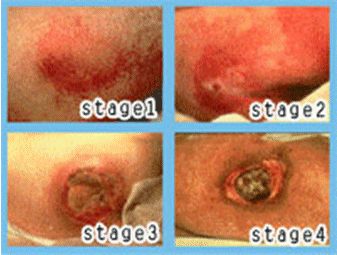
Pressure sores are grouped by their severity. Stage I is the earliest stage. Stage IV is the worst.
  • Stage I: A reddened area on the skin that, when pressed, does not turn white. This is a sign that a pressure ulcer is starting to develop.
  • Stage II: The skin blisters or forms an open sore. The area around the sore may be red and irritated.
  • Stage III: The skin now develops an open, sunken hole called a crater. There is damage to the tissue below the skin.
  • Stage IV: The pressure ulcer has become so deep that there is damage to the muscle and bone, and sometimes to tendons and joints.

 
Physical Examination Pressure Ulcers in Elderly

1. General Condition
Generally, patients come to the sick and agitated or anxious as a result of damage to the integrity of the skin is experienced.

2. Vital Signs
Normal blood pressure, rapid pulse, increased temperature and respiration rate increased.

3. Examination of the Head and Neck

a) Head and Hair
Examination includes the shape of the head, deployment and change hair color as well as an examination of the wound. If there is injury to the area, causing pain and skin damage.

b) Eye
Includes symmetry, conjunctiva, pupil reflexes to light and impaired vision.

c) Nose
Includes examination of the nasal mucosa, cleanliness, nostril breathing does not arise, there is no discharge.

d) Oral
Note the presence of cyanosis or condition dry lips.

e) Ear
Note the form of hearing loss due to foreign bodies, bleeding and wax. In patients who bet the rest by his side, is likely to occur in areas of ulcer ears.

f) Neck
Knowing the position of the trachea, carotid pulse, presence or absence of jugular vein and gland enlargement linfe.

4. Examination Chest and Thorax
Inspection form of the thorax and lung expansion, auscultation respiratory rhythm, vocals premitus, the extra noise, heart sounds, and extra heart sounds, percussion thorax to look for abnormalities in the thorax area.

5. Abdomen
Form flat or flat stomach, bowel sounds decrease due to immobilization, there are masses because of constipation, and abdominal percussion hypersonor if abdominal distention or tense.

6. Urogenital
Inspection abnormalities in the perineum. Usually clients with paraplegia ulcers and catheter attached to urinate.

7. Musculoskeletal
A fracture in the bone will cause the client bet rest in a long time, so
a decline in muscle strength.

8. Examination of Neurology
The level of consciousness assessed with GCS system. Value could decrease if there is pain (neurogenic shock) and heat or high fever, nausea, vomiting, and stiff neck.

9. Physical Assessment of Skin

Assessment involves the skin around the area of ​​the skin including the mucous membranes, scalp, hair and nails. Appearance of skin that needs to be assessed is the color, temperature, humidity, dryness, skin texture (rough or smooth), lesion vascularity.
That must be considered by the nurse is:

a) The color, affected by blood flow, oxygenation, temperature and pigment production.
The lesions were divided into two: a) the primary lesion, which occurs due to a change in one component of skin. b) secondary lesions are lesions that appear after the primary lesion. Preview lesions that must be considered by the nurse that the color, shape, location and kofigurasinya.

b) Edema
During the inspection of the skin, the nurse records the location, distribution and color of the region edema.

c) Humidity
Normally, humidity increases due to increased activity or high ambient temperatures of dry skin can be caused by several factors, such as dry or moist environments that are not suitable, inadekuat fluid intake, the aging process.

d) Integrity
That must be considered is the location, shape, color, distribution, if there is drainage or infection.

e) Cleanliness skin

f) vascularization
Bleeding from the blood vessels and produces petechie echimosis.

g) Palpation of skin
To note that the lesions on the skin, moisture, temperature, texture or elasticity, skin turgor.

10. examination Support

1) A complete blood
Certain increase in hemoglobin concentration early show, with respect to the displacement or loss of fluid and to detect nutritional deficiencies clients. If there leukocytosis due to loss of cells in the inflammatory response to injury and edema. Serum glucose increased due to the stress response.

2) Biopsy wound
To determine the number of bacteria.

3) Swab culture
To identify the type of bacteria on the surface of the ulcer.

4) Preparation of clinical pictures
Created to demonstrate the nature and extent of skin disorders or ulcers and used for improvement after therapy.

Signs and Symptoms of Alzheimer's Disease

Signs and Symptoms of Alzheimer's Disease
Alzheimer's Disease

 
1. Activity / rest
Symptoms: feeling tired
Signs: day / night anxiety, helplessness, sleep pattern disturbance
Lethargy: decreased interest or concern to the usual activities, hobbies, inability to mention again what is read / follow event television programs.
Impaired motor skills, inability to perform usual things have been done, which is very useful movement.

2. Circulation
Symptoms: History of cerebral vascular disease / systemic hypertension, embolic episodes (a predisposing factor).

3. Ego integrity
Symptoms: Suspicious or afraid of the situation / person fantasies, misperceptions of the environment, fault identification of objects and people, hoarding objects: believing that the wrong object placement, has been stolen. Loss of multiple, changes in body image and self-esteem perceived.
Signs: Hiding disability (many reasons are unable to perform an obligation, it may also open the hand without reading the book yet), sit back and watch the others, the first activity may accumulate fixtures and emotionally stable, repetitive movements (folding, unfolding, refolding , cloth), hid the goods, or take a walk.

4. Elimination
Symptoms: The urge to urinate
Signs: Incontinence of urine / feaces, tends to constipation.

5. Food / fluid
Symptoms: History of episodes of hypoglycemia (a predisposing factor) changes in taste, appetite, weight loss, denying the hunger / need to eat.
Symptoms: Loss of ability to chew, avoiding / refusing to eat (probably trying to hide skill), and it looks even thinner (advanced stage).

6. Hiygene
Symptoms: Need help / dependent people
Signs: not able to maintain the appearance, personal habits is lacking, poor cleaning habits, forgot to go to the bathroom, forgetting the steps to pee, can not find the bathroom and less interested in or have forgotten at meal time: depending on others to cook and prepare food on the table, eating, using cutlery.

7. Neuro-sensory
Symptoms: The denial of existing symptoms, especially cognitive changes, and or a vague, hypochondriac complaints about fatigue, dizziness or headache sometimes. complaints in cognitive abilities, decision-making, given the past, a decrease in behavior (observed by people nearby). Loss of sensation propriosepsi (position of the body or particular parts of the body in space). and a history of cerebral vascular disease / systemic embolism or hypoxia held periodically (as a predisposing factor), and seizure activity (secondary to brain damage).
Symptoms: Damage communication: aphasia and dysphasia; difficulty in finding the right words (especially nouns); asked repeatedly or conversations with the substance of the word that has no meaning; fragmented, or speech not audible. Losing the ability to read and write stages (loss of fine motor skills).

8. Comfort
Symptoms: A history of serious head trauma (may be a predisposing factor or acceleration factor), traumatic accidents (falls, burns, and so on).
Sign: ecchymoses, lacerations and hostile / attack others

9. Social interactions
Symptoms: Feeling lost power. psychosocial factors previously; personal and individual influences that appear to change patterns of behavior emerge.
Signs: Loss of social control, improper behavior.

NANDA Nursing

Nursing Care Plan