Nursing Diagnosis for Hydatidiform Mole
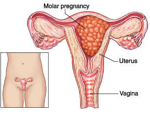
A hydatidiform mole is a relatively rare condition in which tissue around a fertilized egg that normally would have developed into the placenta instead develops as an abnormal cluster of cells. A molar pregnancy is a gestational trophoblastic disease that grows into a mass in the uterus that has swollen chorionic villi. These villi grow in clusters that resemble grapes.
Causes of Hydatidiform Mole
The specific cause behind the occurrence of Hydatidiform mole still remains to be found out. Nevertheless, the doctors point at some reasons like abnormalities in the egg, nutritional deficiencies during pregnancy. If a woman follows a diet which is low in protein, carotene and folic acid she can also contract this malady.
Symptoms of Hydatidiform Mole
- Abnormal growth of the womb (uterus) ; Excessive growth in about half of cases, Smaller-than-expected growth in about a third of cases
- Nausea and vomiting that may be severe enough to require a hospital stay
- Vaginal bleeding in pregnancy during the first 3 months of pregnancy
- Symptoms of hyperthyroidism ; Heat intolerance, Loose stools, Rapid heart rate, Restlessness, nervousness, Skin warmer and more moist than usual, Trembling hands, Unexplained weight loss
- Symptoms similar to preeclampsia that occur in the 1st trimester or early 2nd trimester -- this is almost always a sign of a hydatidiform mole, because preeclampsia is extremely rare this early in a normal pregnancy ; High blood pressure, Swelling in feet, ankles, legs
Diagnosis of Hydatidiform Mole
The physician may not suspect a molar pregnancy until after the third month or later, when the absence of a fetal heartbeat together with bleeding and severe nausea and vomiting indicates something is amiss.
First, the physician will examine the woman's abdomen, feeling for any strange lumps or abnormalities in the uterus. A tubal pregnancy, which can be life threatening if not treated, will be ruled out. Then the physician will check the levels of human chorionic gonadotropin (hCG), a hormone that is normally produced by a placenta or a mole. Abnormally high levels of hCG together with the symptoms of vaginal bleeding, lack of fetal heartbeat, and an unusually large uterus all indicate a molar pregnancy. An ultrasound of the uterus to make sure there is no living fetus will confirm the diagnosis.
Treatment of Hydatidiform Mole
Hydatidiform moles should be treated by evacuating the uterus by uterine suction or by surgical curettage as soon as possible after diagnosis, in order to avoid the risks of choriocarcinoma.[19] Patients are followed up until their serum human chorionic gonadotrophin (hCG) level has fallen to an undetectable level. Invasive or metastatic moles (cancer) may require chemotherapy and often respond well to methotrexate. The response to treatment is nearly 100%. Patients are advised not to conceive for one year after a molar pregnancy. The chances of having another molar pregnancy are approximately 1%.
Management is more complicated when the mole occurs together with one or more normal fetuses.
Carboprost (PGF2α) medication may be used to contract the uterus.
Nursing Diagnosis for Hydatidiform Mole
1. Acute Pain
2. Activity Intolerance
3. Disturbed Sleep Pattern
4. Hyperthermia
5. Anxiety

0 komentar: